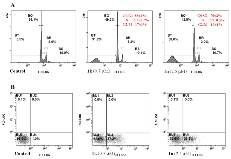
Figure 3.
Effect of compounds 1k and 1n on cell cycle distribution (A) and PS externalization of MCF-7 cells (B). Cell monolayers were incubated for 24 h in the absence (control) or in the presence of individual compounds and submitted to flow cytometric analysis after propidium iodide (A) or AnnexinV/PI double staining (B) as reported in 3.2 Paragraph. (A) Percentage of viable cells in the different phases are given in each picture. Values are the mean ± SD of three separate experiments in triplicate. Representative images of three experiments with comparable results; (B) BU3, viable cells (AnnexinV−/PI−); BU4, cells in early apoptosis (AnnexinV+/PI−); BU2, cells in tardive apoptosis (AnnexinV+/PI+); BU1, necrotic cells (AnnexinV−/PI+). Representative images of three experiments with comparable results.

