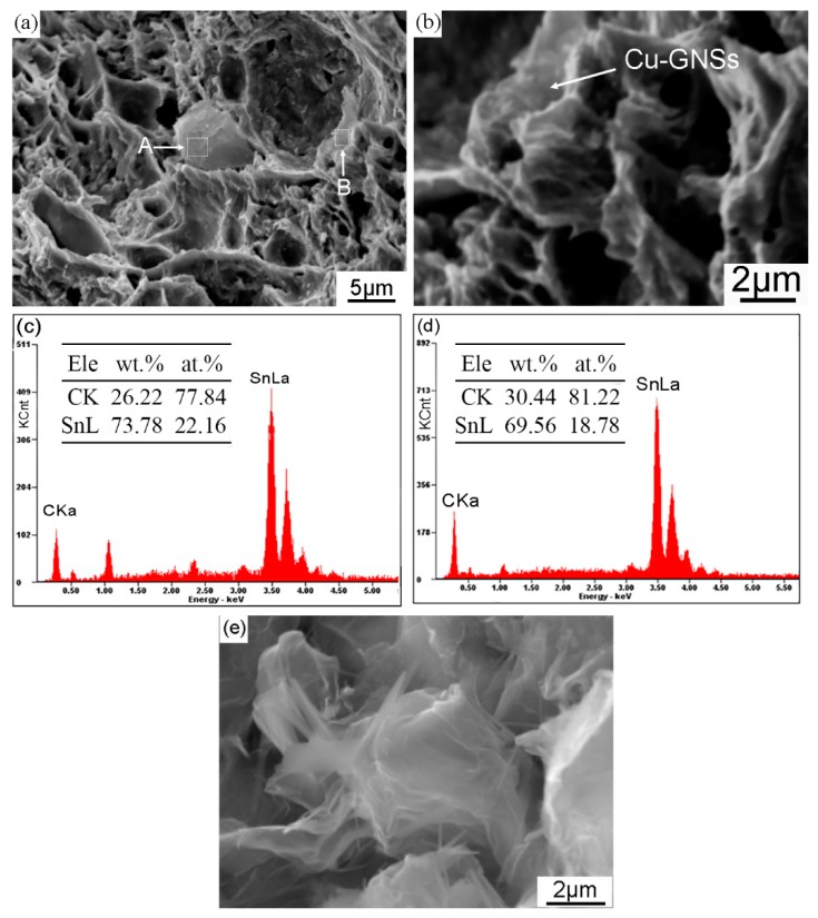
Figure 5.
Scanning electron microscope micrographs of: (a) tensile fractograph of composite solder at 0.05 wt.% Cu-GNSs; (b) high magnification image of region B with Cu-GNSs clung to the edge of the dimple; (c,d) corresponding EDS spectra taken from the white rectangles marked as A and B in (a), respectively; (e) raw material of GNSs.

