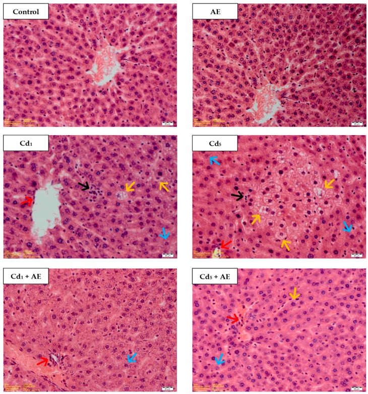
Figure 7.
The effect of the extract from the berries of Aronia melanocarpa L. (AE) on the histological structure of the liver of rats exposed to cadmium (Cd) for 24 months. The rats received Cd in diet at the concentration of 0, 1, and 5 mg Cd/kg and/or 0.1% aqueous AE or not (H + E staining; ×400). Representative sections of the liver are presented. Control: normal liver in the control rats; AE: normal liver in the rats receiving 0.1% AE alone; Cd1 group: blurred trabecular structure—in some lobes, vacuolization and enlarged cells dimensions—in some lobes (↑), microvacuolar steatosis—in some lobes (↑), colliquative necrosis—in some lobes (↑), mononuclear cell infiltrations—in some lobes (↑); Cd5 group: blurred trabecular structure—in some lobes, vacuolization—in majority of cells, in some lobes (↑), colliquative necrosis—in some lobes (↑), microvacuolar steatosis—in some lobes (↑), mononuclear cell infiltrations—in some lobes, (↑); Cd1 + AE group: blurred trabecular structure, vacuolization and enlarged cells dimensions—in some cells in lobes (↑), mononuclear cell infiltrations (↑); Cd5 + AE group: blurred trabecular structure—in some lobes, microvacuolar steatosis—in some lobes (↑), vacuolization—in some lobes (↑), mononuclear cell infiltrations (↑).

