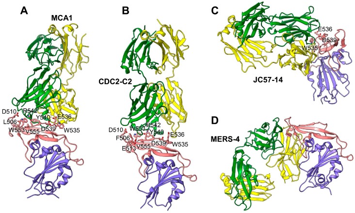
Figure 4.
Structural basis for MERS-CoV RBD recognition by neutralizing mAbs. (A) Crystal structure of MERS-CoV RBD complexed with MCA1 mAb (PDB ID: 5GMQ). (B) Crystal structure of the MERS-CoV RBD complexed with CDC2-C2 mAb (PDB ID: 6C6Z). (C) Crystal structure of the MERS-CoV RBD complexed with JC57-14 mAb (PDB ID: 6C6Y). (D) Crystal structure of MERS-CoV RBD complexed with MERS-4 mAb (PDB ID: 5ZXV). The MERS-CoV RBD core is colored in blue, the RBM is colored in red, and the heavy chains and light chains of each mAb are colored in green and yellow, respectively. The DPP4-binding residues that are blocked by each mAb are shown as sticks. mAb, monoclonal antibody; MERS-CoV, Middle East respiratory syndrome coronavirus; PDB, protein data bank; RBD, receptor-binding domain; RBM, receptor-binding motif.

