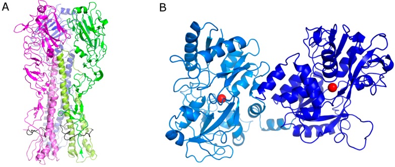
Figure 1.
(A) Cartoon representation of the hemagglutinin (HA) trimeric protein. Each monomer has a different color. The HA1 chain has been depicted with a dark color and HA2 with a pale color. The fusion peptide is represented in black. (B) Cartoon representation of bovine lactoferrin (bLf). The N-lobe is represented in pale blue and the C-lobe in blue. The iron ions are depicted as red spheres.

