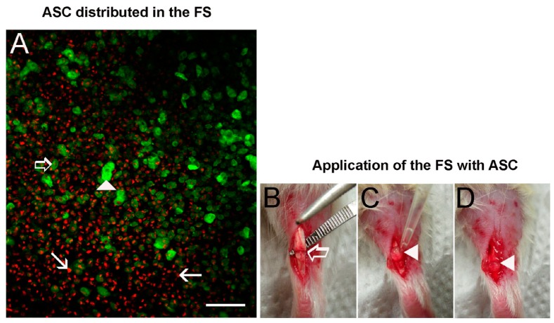
Figure 2.
(A) Confocal microscope image of ASC labeled with phalloidin-FITC (actin cytoskeleton in green) and DAPI (nucleus in red) distributed in the dense fibrin network formed by the FS (n = 3). Observe the disposition of 3.7 × 10⁵ cells in the FS before application in the tendon transected region. (▶) superficial cells, (⇨) intermediately positioned cells and cells at the bottom (→). (B) Model of tendon injury showing the partial transection (⇨) in the proximal region of the Achilles tendon. (C) Application of the FS with ASC using a pipette: note the formation of a clot (▶). (D) Representation of the FS with ASC (▶) covering the transected region before the skin suture. Bar = 200 μm.

