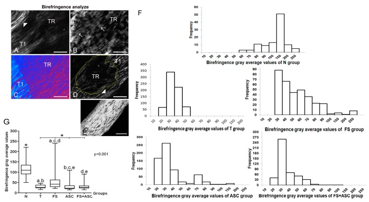
Figure 8.
Images of birefringence of tendon longitudinal sections on 21st day using polarization microscopy (n = 5). The larger tendon axis was set 45° from the crossed polarizers. The variation of brightness intensity (gray levels) is due the variation of the collagen bundles organization. (A) T group: little birefringence brightness is observed in TR (tendon transected region) because of the disorganization of collagen bundles. T1 is the region which border the TR. (▶) remaining portion of the tendon located below the TR. (B) FS group: the increase in birefringence brightness was remarkable and a typical well-developed crimp (→) pattern was observed only in this group. (C) ASC group: image using DIC (differential interference contrast microscopy), where it is possible to visualize in red a smaller organization of the collagen bundles and in intense blue (according to Michel-Lévy’s table) the high degree of compaction of the collagen bundles. (D) FS + ASC group: observe a higher birefringence of the collagen fibers compared to the ASC group and observe an imbrication between collagen fibers that were not cut in T1 and fibrils in the TR (delimited by yellow line). (▶) remaining portion of the tendon located below the TR. (E) N group: collagen fibers exhibiting strong birefringence. Bar = 100 μm (A, C, D, E) and bar = 200 μm (B, F) Frequency histograms of birefringence gray average (GA) values expressed in pixels in the groups N, T, FS, ASC and FS + ASC, which reflect the variability of the collagen fibers organization on the TR region of the Achilles tendon. (G) Birefringence GA (pixels) median between the groups. The measurements data showed in graphics (f and g) were obtained with the larger tendon axis positioned at 45° from the crossed polarizers. (*) Significant differences between the group N and groups with transected tendons. The same letters between the groups correspond to a significant difference between them.

