
Figure 2 – Highlights of proteins with high-confidence assignment of D-methylation or N-deamidation (→methylation) in human RBCs.
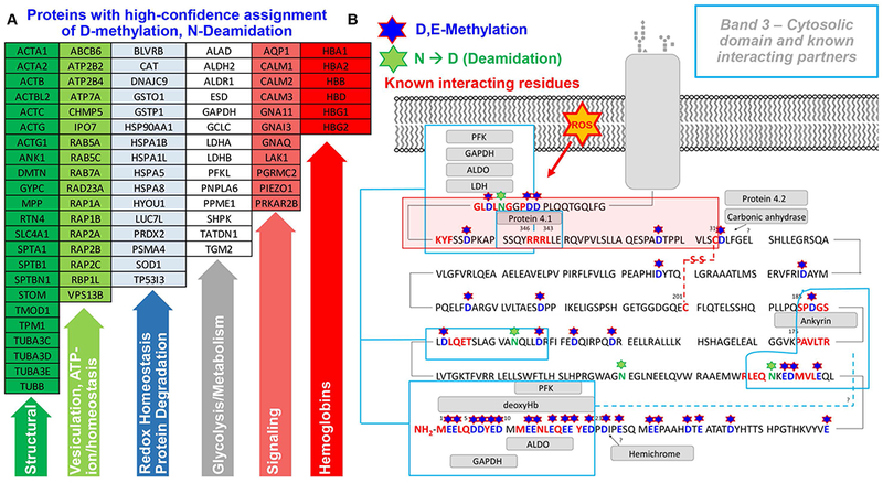
(UniProt abbreviations are listed – A; full list is provided in Supplementary Table 2, along with peptide sequences). In B, an overview of the cytosolic domain of band 3, from residues N-term (1) through residue 380 (SLC4A1_HUMAN; P02730). Stars highlight methylated aspartate and glutamate residues (single letter code D and E, respectively – in blue) as well as deamidated and deamidated → methylated residues (N – in green) on the N-term cytosolic domain of band 3. Rectangles also show the sequences on the N-term of band 3 that have been previously identified54–58 as essential mediators of the interaction between this membrane protein and structural proteins and glycolytic enzymes.