
Figure 4 – Methylation of isoaspartate and deamidated asparagine residues in human hemoglobin alpha and beta,
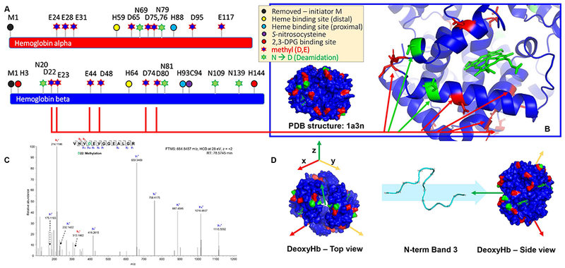
as mapped on their sequence (A), highlighted in the deoxyhemoglobin structure (based on pdb: 1a3n – B) and shown in a representative MS/MS spectrum (methylation of D22 – C). A top and side view of deoxyhemoglobin, along with the N-term of band 3 that has been shown to interact with the deoxyhemoglobin tetramer is shown in D. D-methyl and N-deamidated residues are highlighted in red and green, respectively.