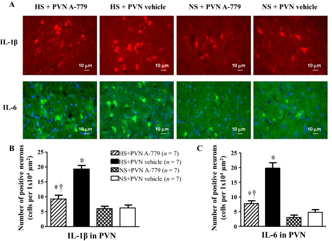
Fig. 3.
Effect of microinjection of A-779 into the PVN on interleukin-1β (IL-1β) and interleukin-6 (IL-6) in the PVN. Immunofluorescence was used to detect pro-inflammatory cytokines in the PVN in different groups. A IL-1β (bright red) and IL-6 (bright green) were both found in the PVN in the different groups. Nuclei were labeled with DAPI (blue). B, C Histograms showing the numbers of IL-1β-positive (B) and IL-6-positive neurons (C) in the PVN of rats in different groups. Values are presented as mean ± SEM; *P < 0.05 versus NS groups (NS + PVN A-779 or NS + PVN vehicle); †P < 0.05 HS + PVN A-779 versus HS + PVN vehicle.

