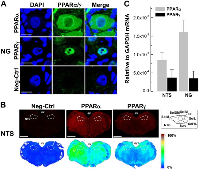
Fig. 1.
Distribution of PPAR-α/PPAR-γ mRNA and protein on the NG and NTS. A Immunostaining of PPAR-α/PPAR-γ protein in NG tissue sections (7 μm). DAPI staining indicates nuclei (blue). Scale bars, 50 μm; n = 6 duplications. B Immunostaining of whole brainstem sections (35 μm; bregma, −12.60 mm) showing the distribution of PPAR-α/PPAR-γ protein in the NTS region. Scale bars, 2 mm; n = 6 duplications. C Levels of mRNA expression of PPAR-α/PPAR-γ in the NTS and NG of normal rats. n = 6 duplications. The condition with no primary antibody was set as the negative control. 4 V, 4th ventricle; Sol, solitary tract; SolL, lateral Sol; SolVL, ventrolateral Sol; SolV, ventral Sol; SolM, medial Sol; SolDM, dorsomedial Sol; SolIM, intermediate Sol.

