Figure 1.
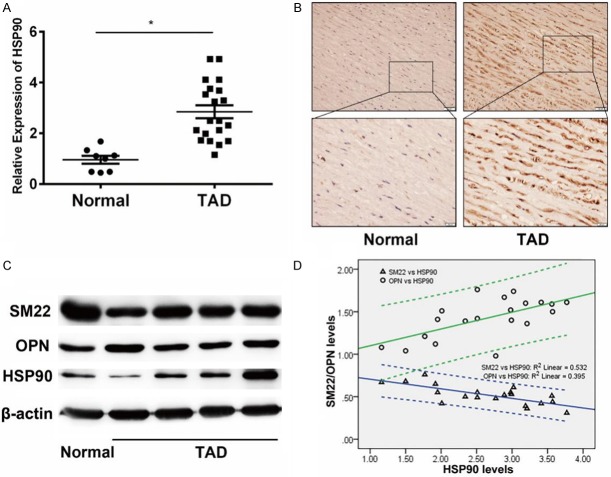
Elevated expression of HSP90 and synthetic phenotypic markers in aortic walls from TAD patients. A. Detection of HSP90 expression in aortic walls from patients with TAD (n=20) and healthy control (n=8) by real-time PCR. The expression of HSP90 in aortic walls from patients with TAD higher than healthy control. β-actin was used as an internal control. *P<0.05 vs Normal group. B. Representative image of immunohistochemistry of HSP90 staining in aortic walls tissues lesions. The staining of HSP90 in aortic walls tissues of TAD patients was higher than normal group. C. Western blot analysis of SM22, OPN and HSP90 expression in aortic walls tissues. Compared with normal group, the expression of HSP90 and OPN were higher in TAD group, while SM22 was lower expressed. D. Spearman analysis of the correlation between protein expression of HSP90 and SMCs phenotypic markers in TAD patients. The protein levels were determined according the densitometry from western blot assay, which was evaluated by Image J software. The expression of HSP90 was positively correlated with OPN while negatively correlated with SM22.

