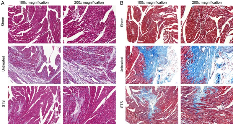
Figure 2.

STS improved myocardial pathological changes in MI mice. A. Representative illustration of hematoxylin and eosin staining of infarcted mouse hearts. These photos demonstrated the intense inflammatory response and myocardial cells arranged irregularly after MI. B. Representative images of Masson’s trichrome-staining infarcted hearts in mice. Blue represents region with replacement fibrosis.
