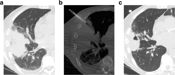
Fig. 1.
a) Axial CT scan in a 69-year-old man status post kidney transplant demonstrates an area of consolidation and surrounding ground glass opacity in the right middle and lower lobe. He underwent bronchoalveolar lavage and transbronchial biopsy which found acute hemorrhage and hemosiderosis. b) Axial CT scan obtained during PTNB, performed 2 days after bronchoscopy, shows the needle within the consolidation in the right middle lobe. Fungal hyphae were identified on cytologic evaluation and aspergillus was identified on accompanying microbiology culture. c) Axial CT performed three months later, on treatment with ambisome, shows the area of consolidation improving

