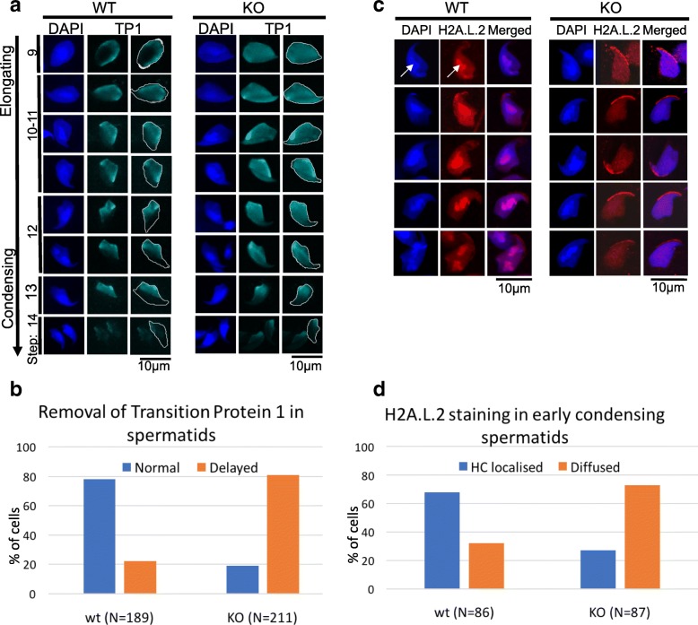
Fig. 4.
Characterization of the incorporation of TP1 and H2A.L.2 into spermatid chromatin in the absence of H2A.B.3. a Elongating/condensing spermatids immunostained with anti-TP1 antibodies (turquoise) and counterstained with DAPI (blue). The outlines of the spermatids nuclei are shown as a white trace in TP1 panels. b The quantification of the number of wild type and H2A.B.3−/y step 10–12 spermatids with a delayed wave of TP1 release. c HA.L.2 localization in condensing wild type and H2A.B.3−/y spermatid. White arrows point to pericentric heterochromatin. d The quantification of the number of wild type and H2A.B.3−/y condensing spermatids with H2A.L.2 enriched in pericentric heterochromatin (HC)

