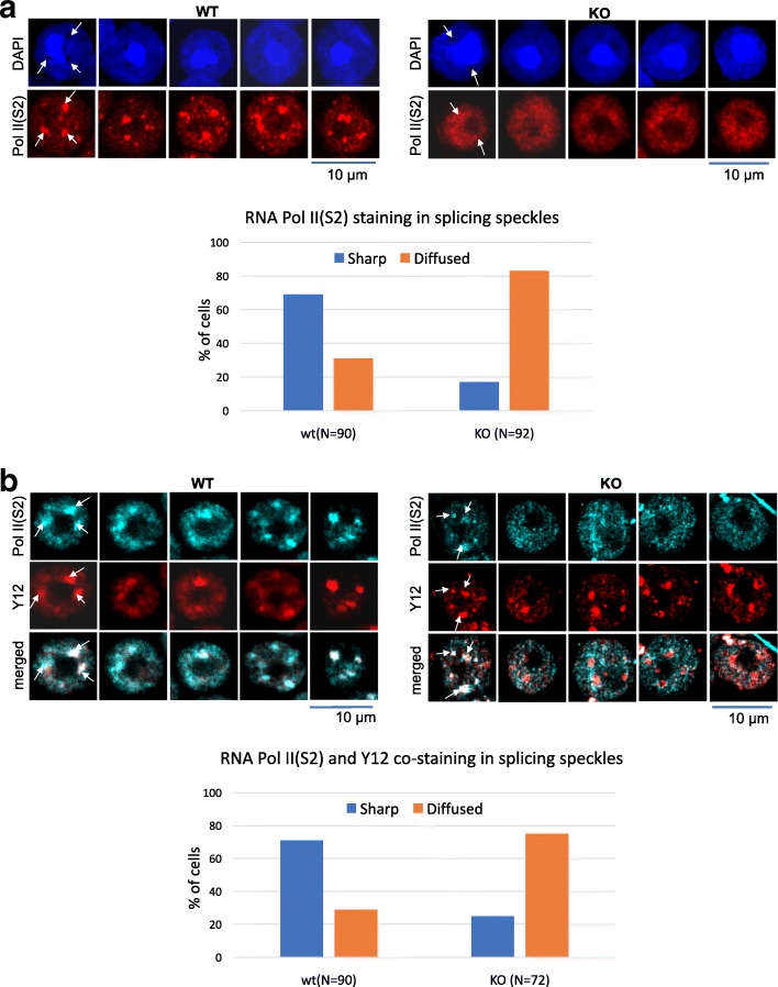
Fig. 6.
Localization of the active form of RNA polymerase II within splicing speckles is altered in H2A.B.3−/y round spermatids. a Immunofluorescence staining of round spermatids from wt and H2A.B.3−/y mice testis. Cells were indirectly labeled with anti-Pol II S2P antibodies and co-stained with DAPI. Scale bar is 10 μm. White arrows show accumulation of RNA Pol II S2P signal in splicing speckles of WT round spermatid cells. The signal is diffused in H2A.B.3−/y. Detailed quantification of RS in wt (n = 90) and H2A.B.3−/y (n = 92) mice showing that majority of RS cells in H2A.B.3 KO mice have diffused Pol II S2 localization. b Immunofluorescence staining of round spermatids from wt and H2A.B.3−/y mice testis. Cells were indirectly labeled with anti-Pol II S2P and Y2 (a splicing speckle marker) antibodies. Scale bar is 10 μm. White arrows show colocalization of RNA Pol II S2P and Y12 in splicing speckles of wt round spermatid cells. RNA Pol II S2P no longer co-stains with Y12 in the majority of round spermatids from H2A.B.3−/y testes. Detailed quantification of RS in wt (n = 90) and H2A.B.3−/y (n = 72) `

