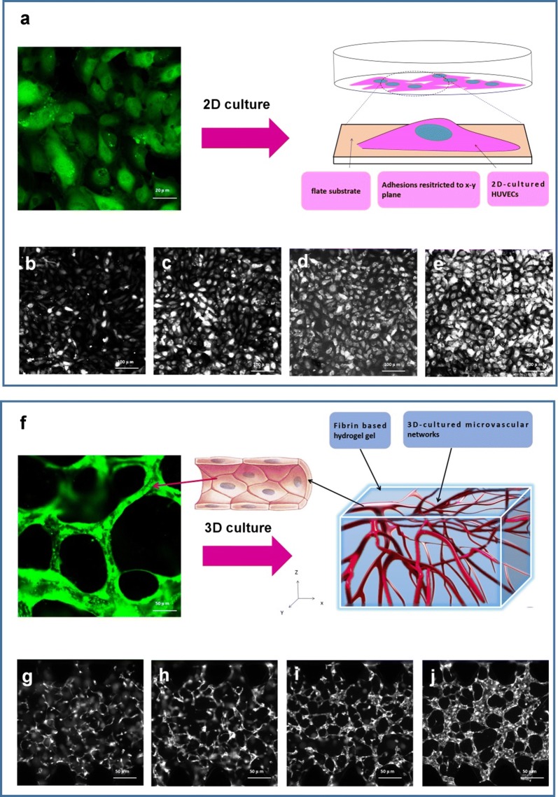
Fig. 3.
Development progress of human umbilical vein endothelial cells (HUVECs) by 2D culture and 3D culture. a 2D HUVEC culture in a disk, b–e camera image of fluorescent HUVECs developed from day 1 to day 4. f 3D HUVEC culture in a chip, g–j camera image of fluorescent HUVECs developed from day 1 to day 4

