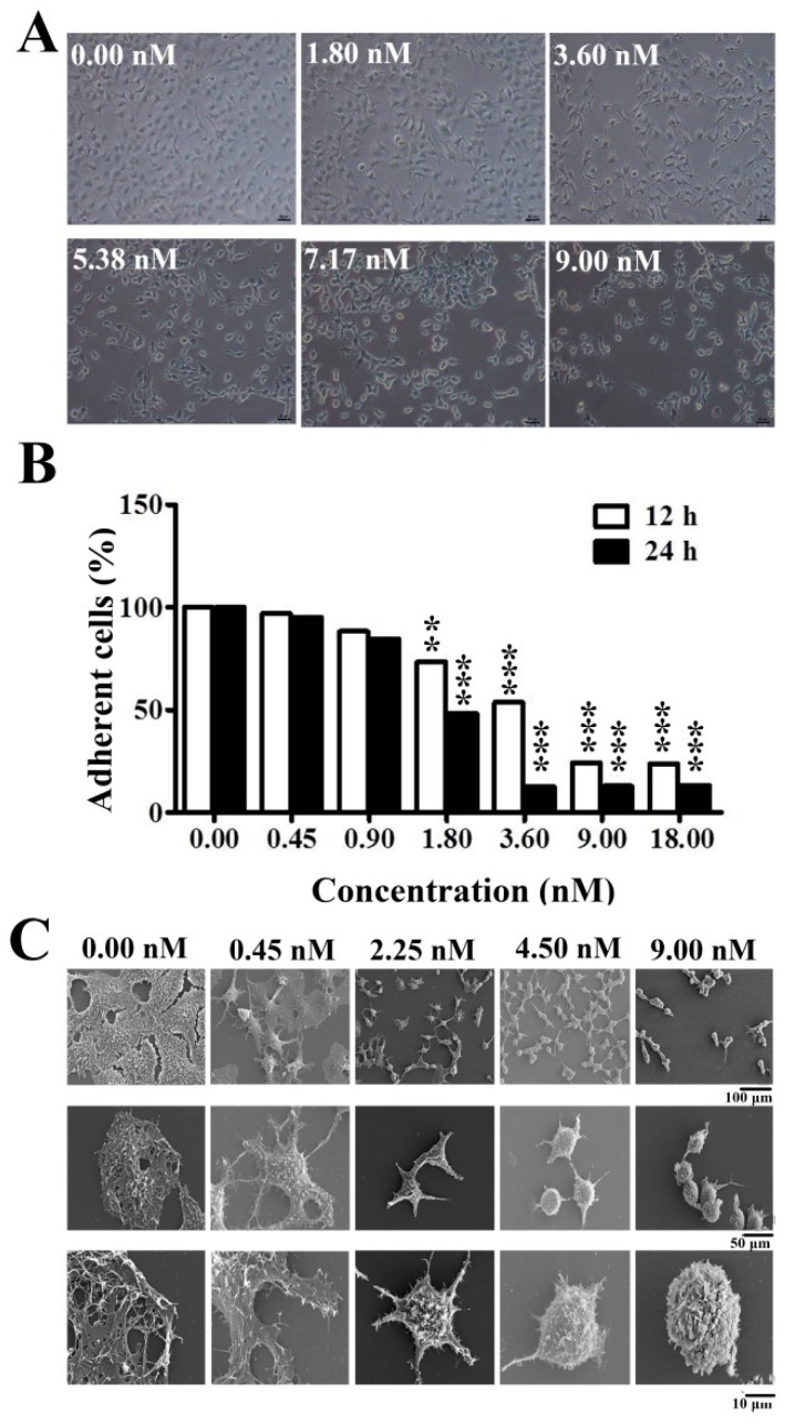
Figure 2.
Inhibition of cell adhesion and destroying of filiform structures in Huh7.5 cells treated by EPS11. (A) Observation of the morphological changes in Huh7.5 cells after the treatment of different concentrations of EPS11 for 6 hours via light microscope (Nikon, Tokyo, Japan). (B) Quantification assay of cell adhesion in Huh7.5 after treatment with different concentrations of EPS11 for 12 hours and 24 hours. The data were presented as means ±SD of three observation fields in one representative experiment chosen from three independent experiments. *p < 0.05, **p < 0.01, ***p < 0.001. (C) Observation of the filiform structures in Huh7.5 cells after the treatment of different concentrations of EPS11 via scanning electron microscopy (SEM). Huh7.5 cells were treated with indicated concentration of EPS11 (0, 2.25, 4.50, 9.00 nM) for 6 hours.

