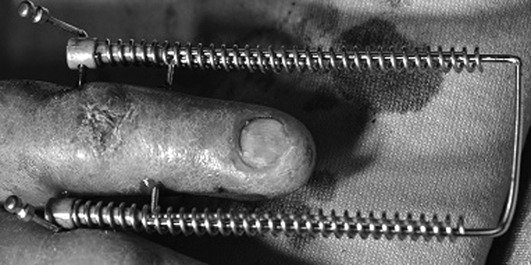
Figure 2.

Clinical photograph of fifth finger, left hand, after Ligamentotaxor® application for PIP joint fracture-dislocation. The placement of the transverse Kirschner wires is made under fluoroscopy with the aid of a guiding device. After placing and modelling the K wires, spherical joints are placed and the metallic springs are screwed. The distraction obtained is controlled under fluoroscopy
