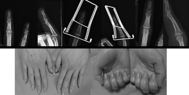
Figure 3.

Clinical case n°1: The figure shows on top, from left to right, the trauma x-rays showing a Seno type 1 fracture of the ring finger (left), post-operative x-rays showing the Ligamentotaxor® in place with the amount of distraction and reduction applied (center) and follow-up x-rays at 4 months, showing fracture healing and initial joint remodelling (right). On the bottom the clinical outcome with ROM in extension (left) and flexion (right)
