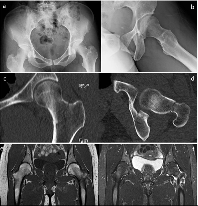
Figure 5.

Case 3. Radiographic images at initial presentation, showing a Type II, compression-side FNSF: (a) panoramic anteroposterior view of the pelvis and (b) lateral view of left hip. Hip CT performed during hospitalisation, (c) coronal and (d) axial views. Pelvis MRI at the same period: T1 image (e) and T2 weighted image (f)
