Figure 1.

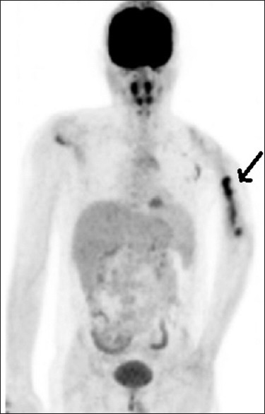
Maximum intensity projection image of fluorodeoxy glucose scan with fluorodeoxy glucose-avid lesion in the left humerus marked by a thick arrow, while the skull lesion cannot be appreciated due to high adjacent normal brain activity

Maximum intensity projection image of fluorodeoxy glucose scan with fluorodeoxy glucose-avid lesion in the left humerus marked by a thick arrow, while the skull lesion cannot be appreciated due to high adjacent normal brain activity