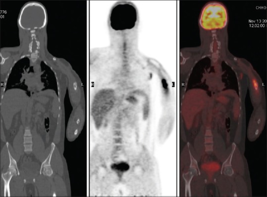
Figure 2.

Coronal sections of computed tomography, positron emission tomography, and fused positron emission tomography/computed tomography images, respectively, showing an increased fluorodeoxy glucose uptake in lytic-sclerotic lesion of the left humerus
