
Fig. 5. 3D visualization of the vasculature in the mouse brain and kidney after FDISCO clearing.
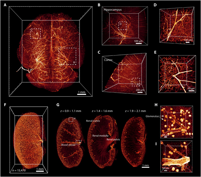
The vasculature was labeled by injection of CD31-A647 antibody. (A) 3D reconstruction of the vasculature in the whole brain after FDISCO clearing and LSFM imaging. (B to E) The details of blood vessels in the hippocampus (B) and cortex (C) are shown. High-magnification views of the dashed boxed regions in (B) and (C) are shown in (D) and (E), respectively. (F) 3D reconstruction of blood vessels and glomeruli in the kidney. The number of glomeruli was counted as 15,470 by Imaris software. (G) Images at gradient depth. The glomeruli were mainly distributed in the renal cortex. (H and I) High-magnification views of the dashed boxed regions in (G).