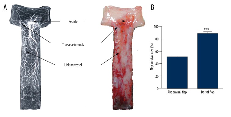
Figure 5.
(A) Dorsal reticulate vessel flap (left: perfusion X-ray films; right: perfusion specimens). The proximal flap containing definite true anastomosis and direct linking vessels survived and the distal flaps were partly necrotic. (B) Quantification of the flap survival area of the different flap types. *** P<0.001.

