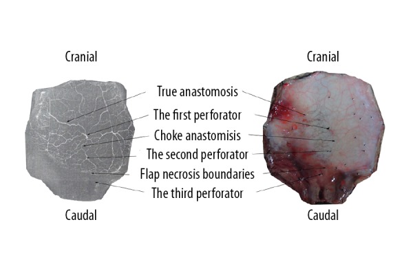
Figure 6.

Single perforator flap (left: perfusion X-ray films; right: perfusion specimens). The flap survived completely in the area with an adjacent perforator (the second perforator) but was partly necrotic in the area with 2 adjacent consecutive perforators (the third perforator). The flap necrosis boundaries were located between the second perforator and the third perforator, and the surviving area of the flap was round in shape.
