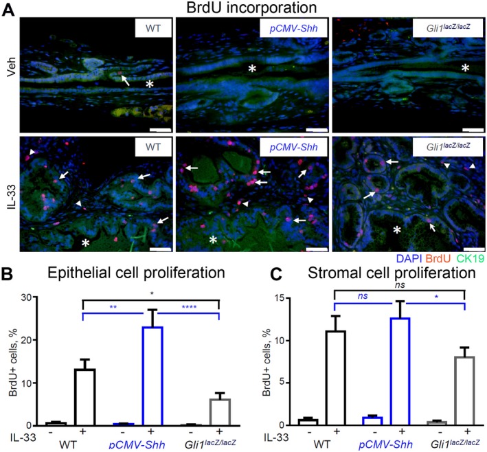
Figure 5.

HH signaling synergizes with IL‐33 to promote biliary cell proliferation. EHBDs from the WT, pCMV‐Shh, and Gli1lacZ/lacZ mice treated with either Veh or IL‐33 (1 µg/mouse/day for 4 days; necropsy on day 5) were analyzed with immunofluorescence for cell proliferation. (A) Proliferating cells were labeled by BrdU incorporation (50 mg/kg, intraperitoneally, 2 hours before necropsy) (red). Cell nuclei were marked with DAPI (blue) and BECs with CK19 (green; original magnification ×400). (B,C) Proliferating cells were quantified in the epithelial (arrows) and stromal (arrowheads) cell compartments with ImageJ software (n = 5‐6 animals per group; five or more high‐power fields per BD; >500 cells/animal). Results are expressed as mean ± SEM; one‐way ANOVA; *P < 0.05, **P < 0.01, ****P < 0.0001. Abbreviation: ns, not significant.
