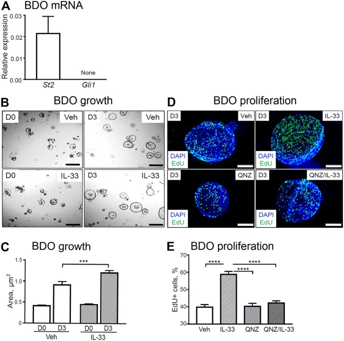
Figure 7.

IL‐33‐induced BDO cell proliferation in vitro. (A) Total RNA was isolated from the WT mouse‐derived organoids and analyzed for mRNA expression of St2 and Gli1 (n = 3 organoid lines). (B,C) BDOs were treated with Veh or recombinant IL‐33 (100 ng/mL) for 72 hours (day 3) starting 24 hours after passage of established BDOs and analyzed for growth (n = 6 technical replicates per group). (D,E) In a separate experiment, BDOs were treated with either Veh or pretreated for 1 hour with the NF‐κB inhibitor QNZ (1 µM) and then treated with recombinant IL‐33 (100 ng/mL) for 72 hours (day 3) after passaging of established BDOs. BDOs were analyzed for proliferation (n = 3 technical replicates per group). (D) The organoids were incubated with EdU for 9 hours to label proliferating cells (green) among all BDO cells (cell nuclei, DAPI [blue]; original magnification ×600). (E) ImageJ software was used to enumerate proliferating cells. Results are expressed as mean ± SEM; one‐way ANOVA (C,E); ***P < 0.001, ****P < 0.0001. Abbreviation: D0/D3, day 0/day 3.
