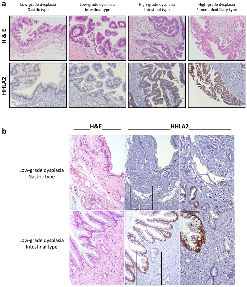
Fig. 3. Prevalent upregulation of HHLA2 expression in IPMN but not in adjacent tissue.
(a) Representative specimens showing HHLA2 experssion pattern in IPMN lesions with low-grade or high-grade dysplasia and different morphological subtypes. H&E staining and HHLA2 IHC from the same section of the TMA cores are presented. Magnification: 200x. (b) Representative IPMN specimens showing variable HHLA2 IHC staining, with only rare scattered proliferating HHLA2 positive ductules within adjacent chronic pancreatitis. H&E staining and HHLA2 IHC from the same section of the TMA cores are presented. The low grade gastric type IPMN is negative for HHLA2; within the adjacent chronic pancreatitis, scattered proliferating ducts are HHLA2 positive. The low grade intestinal type IPMN is positive for HHLA2; within the adjacent chronic pancreatitis, pancreatic ducts are negative for HHLA2. Magnification: 200x, 400x (inset).

