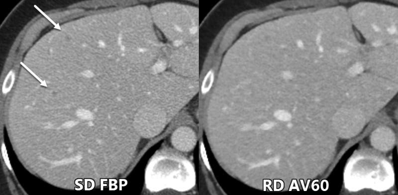
Figure 3:

Axial contrast-enhanced CT images show example of two small low-contrast liver metastases (arrows) that were seen by all three readers during the evaluation of standard radiation dose (SD) filtered back projection (FBP) images but were missed during the evaluation of reduced dose (RD) adaptive statistical iterative reconstruction–V 60% (AV60) images.
