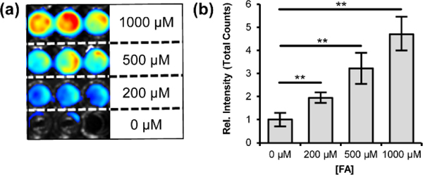
Figure 2.

Chemiluminescence detection of FA in live cells with CFAP540. HEK293 cells were treated with CFAP540 (10 μM, 0.1% DMSO) and incubated for 30 minutes in growth medium. Growth medium was removed and cells were washed thrice with PBS (pH 7.4) Then, cells were treated with 0, 200, 500 and 1000 μM formaldehyde and the chemiluminescence signal was collected for 90 minutes. (a) Representative chemiluminescence cell images in triplicate. (b) Quantification of total light emitted from the cells after 90 minutes. Error bars are ±SEM (n = 6), and statistical analyses were performed with a two-tailed Student’s t test where **P ≤ 0.01.
