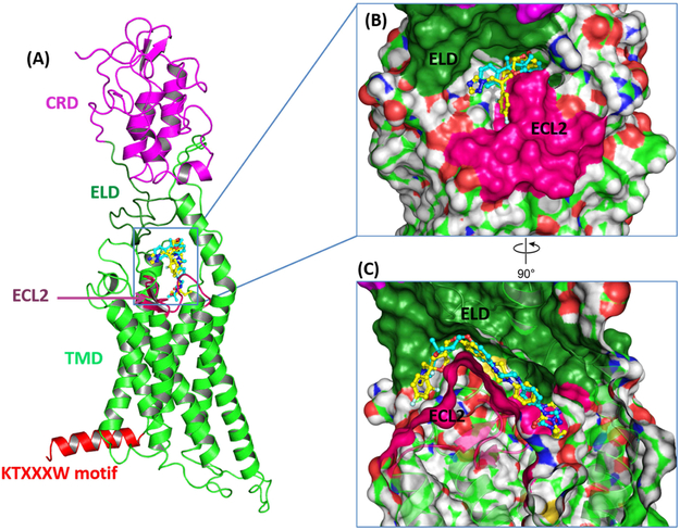
Figure 2.
Binding mode of PSZ and 1 in complex with Smo. (A) The binding orientations of PSZ (yellow) and 1 (blue) inside the binding pocket on Smo. The opening of the binding site is sandwiched between the ELD and ECL2. (B) The furan region of PSZ and 1 orient out of the binding pocket towards the ELD. (C) Compared to PSZ, 1 penetrates more deeply inside the binding pocket.

