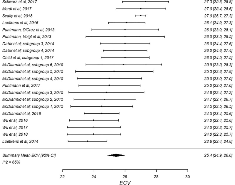
Figure 5a:
Forest plots of extracellular volume (ECV) studies at 3.0-T cardiac MRI in healthy participants. Studies are grouped by vendor and pulse sequence scheme. (a) ECV studies from Philips modified Look-Locker inversion recovery (MOLLI) subgroup and (b) ECV studies from Siemens MOLLI subgroup. Studies with multiple subgroups are noted by author last name and year of publication.

