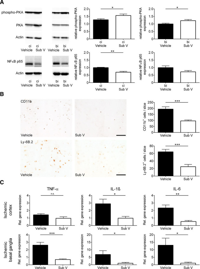
Figure 3.

Substance V (Sub V) exerts anti-inflammatory effects in ischemic stroke. A, Intracellular downstream signaling of cAMP in the ipsilesional cortex (ci) and basal ganglia (bi) of vehicle- and Sub V–treated mice was analyzed by immunoblotting 24 h after transient middle cerebral artery occlusion (tMCAO) using antibodies against NF-κB (nuclear factor-κB) p65 and PKA (protein kinase A). The protein phosphorylation was expressed as a relative amount of phosphoprotein to total protein. Actin was used as loading control. In addition to the densitometric quantification, one representative immunoblot is shown (n=4–5). B, Left, Representative immunocytochemical staining against CD11b+ macrophages/microglia and against Ly-6B.2+ neutrophils in the ischemic hemisphere of vehicle-treated mice and mice treated with 0.5 mg/kg Sub V. Scale bar=100 µm. Right, Quantification of CD11b+ macrophages/microglia and Ly-6B.2+ neutrophils per slice in the infarcted hemispheres on day 1 after tMCAO (n=4). C, Relative gene expression of TNF (tumor necrosis factor)-α, IL (interleukin)-1β, and IL-6 in the ischemic cortices (top) and basal ganglia (bottom) of vehicle- and Sub V–treated mice 24 h after tMCAO (n=4–5). Statistical significances analyzed by (A, B, and C) Student t test and (B; Ly-6B.2+ cells) Mann-Whitney U test. *P<0.05, **P<0.01, and ***P<0.001.
