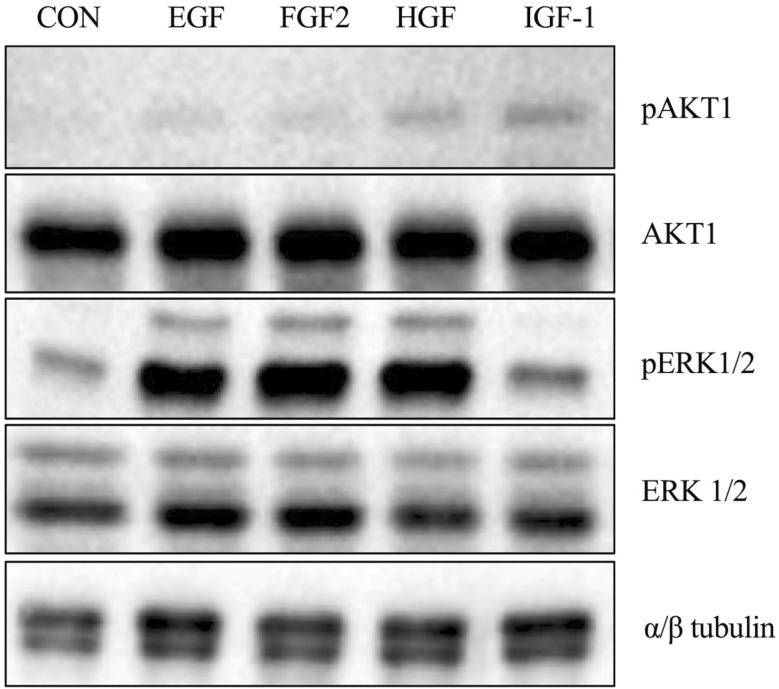
Figure 2.
Phosphorylation of AKT1 and ERK1/2 in response to selected growth factors. Equine iTr cells were serum starved and treated with 10 ng/mL of EGF, FGF2, HGF, or IGF-1 for 20 min prior to lysis. Proteins were separated through SDS-PAGE, transferred to nitrocellulose and analyzed by western blot for total and phosphorylated version of AKT and ERK1/2. Tubulin expression was monitored as a loading control.

