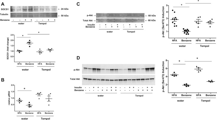
Figure 7.
TEMPOL treatment restores insulin signaling in benzene-exposed mice. Mice drinking normal water or water supplemented with 1 mM TEMPOL were exposed to HFA or 50 ppm benzene for 2 weeks. After euthanasia, the abundance of Socs1 protein was measured in liver homogenates by Western blotting (A; n = 4). Tubulin levels were used as a loading control. Illustrated are representative blots (upper panels) and normalized data (lower panels). Levels of insulin-stimulated phospho-Irs-2 from liver homogenates were also determined using a specific ELISA (B; n = 4). Levels of phospho-Akt and total Akt were determined by Western blotting in homogenates of liver (C; n = 5–10), and skeletal muscle (D; n = 4) from mice variably treated with insulin as indicated. Illustrated are representative blots (left panels) and the calculated induction of insulin-stimulated Akt phosphorylation (right panels); *p < .05.

