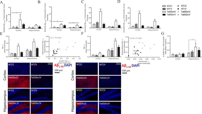
Fig. 5.
Vaccinated Ts65Dn mice exhibit lower levels of cerebral, soluble and insoluble Aβ40/42 and S396-P-tau at 15 m of age. Tissue levels of S396-P-tau protein, soluble and insoluble Aβ40 and Aβ42 were measured quantitatively using sELISA (A-D, G) and IHC (E-F). (A) Cortical levels of soluble Aβ40 were higher in sham-vaccinated Ts65Dn mice compared with both WT mice (P < 0.05). (B) Cortical and hippocampal levels of soluble Aβ42 were higher in sham-vaccinated Ts65Dn mice compared with both WT mice (P < 0.05). (C) Cortical and hippocampal levels of insoluble Aβ40, were higher in sham-vaccinated Ts65Dn mice compared with vaccinated Ts65Dn and WT mice (P < 0.001, P < 0.01, respectively). (D) Cortical and hippocampal levels of insoluble Aβ42 were higher in sham-vaccinated Ts65Dn mice compared with vaccinated Ts65Dn and WT mice (P < 0.001, P < 0.01, respectively). (E) IHC analysis for Aβ40 reveals higher levels in the cortex and hippocampus of sham-vaccinated Ts65Dn mice compared with vaccinated Ts65Dn and WT mice (P < 0.01, upper-left and bottom panels), with high positive correlation between measurements in the cortex and hippocampus (Pearson’s r = 0.7, P < 0.05, upper-right panel). (F) IHC analysis for Aβ42 reveals higher levels in the cortex and hippocampus of sham-vaccinated Ts65Dn mice compared with vaccinated Ts65Dn and WT mice (P < 0.05 for the cortex and P < 0.01 for the hippocampus, upper-left and bottom panels), with medium positive correlation between measurements in the cortex and hippocampus (Pearson’s r = 0.55, P < 0.05, upper-right panel). (G) Hippocampal levels of S396-P-tau protein were higher in sham-vaccinated Ts65Dn mice compared with sham-vaccinated WT mice (P < 0.05), Repeated-measures Two-way ANOVA, Pearson’s correlation coefficient, *P < 0.05, **P < 0.01, ***P < 0.001.

