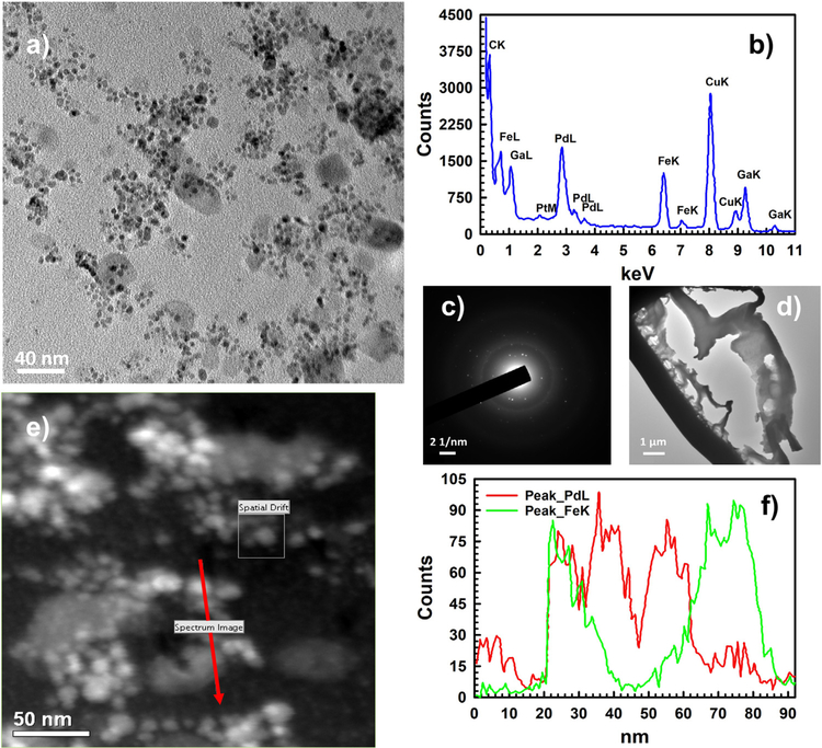
Fig. 9.
Inner surface (a thin lamella was prepared and lifted out from the inside with the help of FIB) of the Pd-Fe-PAA-PVDF membrane (20 wt% monomer and 1 mol% cross-linker) characterized by TEM, HRTEM and selected area electron diffraction (SAED). (a) A typical TEM image of the inner surface showing Pd/Fe nanoparticles (50 K magnification), (b) reproduced EDS spectrum showing peaks of Fe and Pd elements (100 K magnification). During preparation of lamella, gallium was used and signal confirms that. The copper signal is due to the sample holder made of copper, (c) the SAED pattern shows a diffraction halo (representing core carbon of the polymer) and multiple diffraction rings representing different phases of Pd and Fe elements (100 K magnification), (d) the lamella of the inner surface where HRTEM and SAED were conducted (2 K magnification), (e) survey image of inner surface conducted STEM mode (200 K magnification), (f) reproduced EDS signal profile for elemental mapping of the survey image (e) showing presence of Pd and Fe elements distribution in the high-lighted red arrowed line (200 K magnification).

