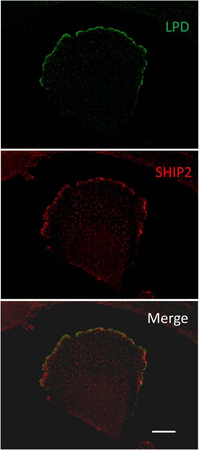
Fig. 4.
SHIP2 and lamellipodin (LPD) colocalization in glioblastoma 1321 N1 cells. 1321 N1 cells were plated on coverslips and kept in culture in the presence of 10% serum for 24 h. The cells were fixed and stained with anti-lamellipodin in green (Alexa Fluor 488) and anti-SHIP2 (Novus) in red (Alexa Fluor 594). Images were obtained on Axioimager (Zeiss) at 100× 1.45 NA oil after deconvolution. Scale bar = 10 µm.

