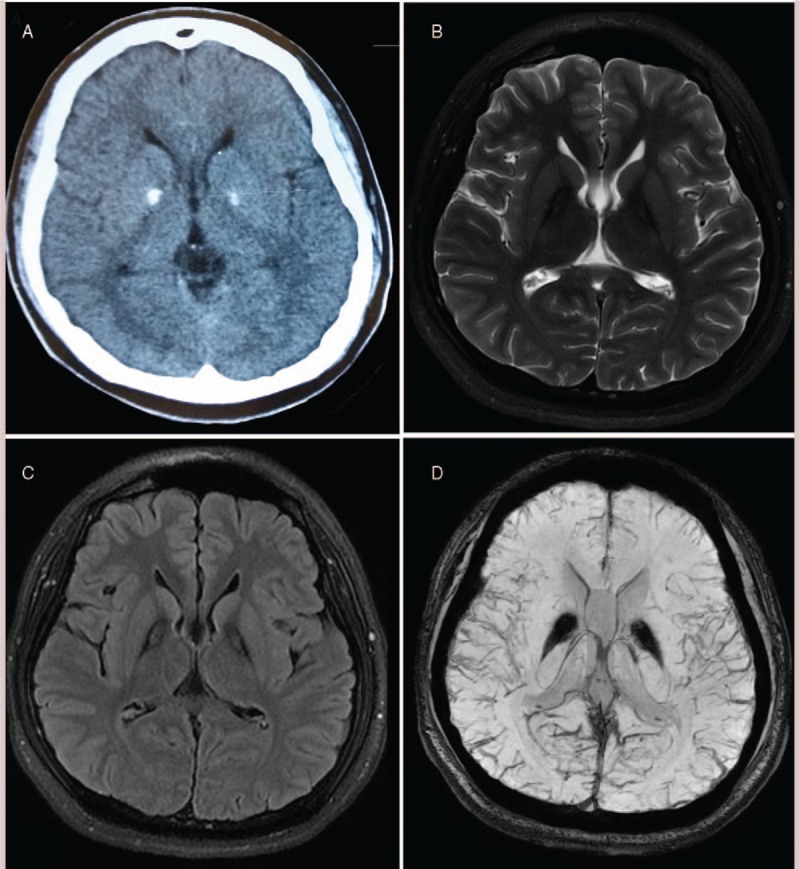
Figure 1.

T2-weighted brain MRI showing the “eye-of-the-tiger” sign. (A) Brain CT scan showed a bilateral calcification of the globus pallidus; (B and C) T2 weighted and FLAIR brain MRI displayed typical “eye of the tiger”; (D) brain SWI showed bilateral symmetrical low signal in the globus pallidus. CT = computed tomography, FLAIR = fluid-attenuated inversion recovery, MRI = magnetic resonance imaging, SWI = susceptibility weighted imaging.
