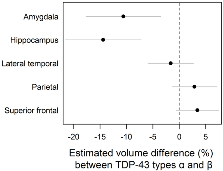
Figure 4.
We investigated the imaging data further by fitting linear regressions adjusting for age at death, total intracranial volume, and Braak stage. Our analysis showed that for cases with TDP-43 type-α had an estimated 11% (4%, 18%, p = 0.003) smaller amygdala volume than TDP-43 type-β. For hippocampi, TDP-43 type-α cases had 14% (7%, 22%, p <0.001) smaller volume than TDP-43 type-β cases. Plotted values are regression estimates and 95% confidence intervals.

