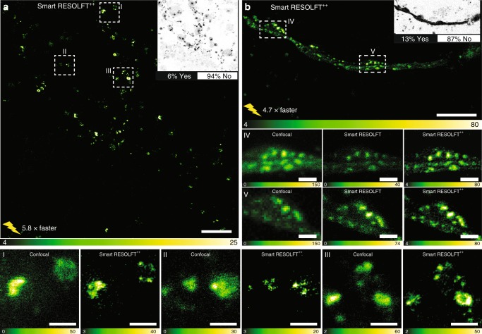
Fig. 4.
Smart RESOLFT imaging of hippocampal neurons. a, b Smart RESOLFT images of Synapsin1A-DronpaM159T, using probe type 2. The images were recorded 5.8 and 4.7 times faster than in conventional RESOLFT imaging with a reduced light dosage of 90% and 83% respectively. The insets show the decision maps with the percentage of skipped pixels (white, NO) and the fully illuminated pixels (black, YES). FOV of 40 by 40 µm2 (a), and 30 by 15 µm2 (b). I−III and V−IV region of interest (ROI) of images (a) and (b) respectively, showing single synaptic vesicles and small clusters. Scale bars are 10 µm (a), 5 µm (b), and 1 µm in ROI I−V. RESOLFT REversible Saturable/Switchable Optical Linear Fluorescence Transitions

