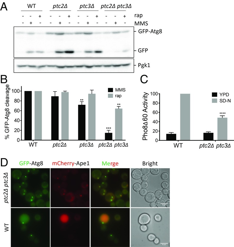
Fig. 1.
Ptc2 and Ptc3 phosphatases redundantly promote rapamycin-induced autophagy. (A) WT and ptc2Δ ptc3Δ strains were grown in YEP-lac until they reached early exponential phase and treated with 200 ng/mL rapamycin or 0.04% MMS for 4 h. Samples were blotted for GFP to quantify GFP-Atg8 cleavage and for Pgk1 as a loading control. (B) GFP-Atg8 cleavage was calculated as the ratio of the free GFP band to the total GFP signal in the lane for three independent experiments and then normalized to WT. Error bars represent SE (**P < 0.002 and ***P < 0.0002). (C) Pho8Δ60 activity in WT cells and the phosphatase-null strain 4 h after nitrogen starvation, normalized to the WT starvation conditions for four independent experiments. Error bars represent SE (****P < 0.0001). (D) Localization of GFP-Atg8 and mCherry-Ape1 in WT and ptc2Δ ptc3Δ at 1 h after rapamycin treatment. (Scale bar: 8 μm.)

