Fig. 1.
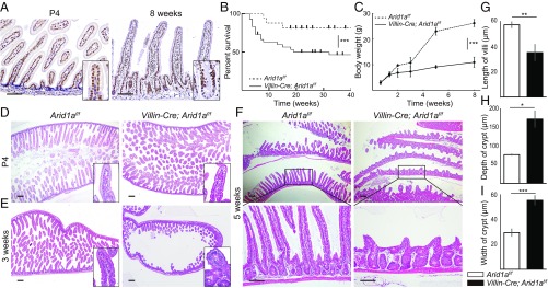
Intestinal Arid1a deletion results in growth impairment, low survival rate, and abnormal intestinal structure in mice. (A) IHC for Arid1a in wild-type mice at P4 (Left) and at 8 wk of age (Right). (B) Kaplan–Meier survival curves show significantly lower survival rate (P < 0.001) in Villin-Cre;Arid1af/f mice (line, n = 30) compared with control mice (dashed line, n = 24). (C) Body weight at indicated time points for control (dashed line, n = 6, male mice) and Villin-Cre; Arid1af/f mice (line, n = 6, male mice). (D–F) H&E staining of the small intestines for control (Left) and Villin-Cre; Arid1af/f mice (Right) at the indicated time points. There was no significant difference between control mice and Villin-Cre;Arid1af/f mice at P4 (D). At 3 wk of age, the intestinal architecture of Villin-Cre;Arid1af/f mice occasionally appeared to be abnormal compared with that in control mice (E). At 5 wk of age, disorganized intestinal architecture, including shortened villi and crypt enlargement, was constantly observed in Villin-Cre;Arid1af/f mice (F). (G–I) Average length of villi (G), depth of crypts (H), and width of crypts (I) in control and Villin-Cre;Arid1af/f mice at 8–10 wk of age (n = 3). [Scale bars, 100 µm (A and F) and 200 µm (D–F).] [Inset magnification, 2.7× (A) and 10× (D and E).] Quantitative data are presented as means ± SD, *P < 0.05, **P < 0.01, ***P < 0.001.

