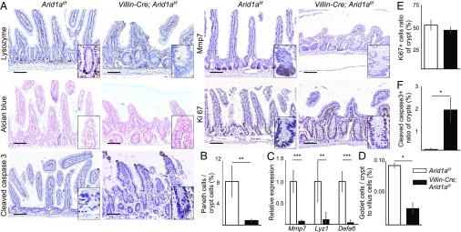
Fig. 2.
Intestinal Arid1a deletion leads to decreased secretory cell lineages and increased apoptotic cells in the small intestines of mice. (A) IHC analysis for Lysozyme, Mmp7, Alcian blue, Ki67, and cleaved caspase 3 staining of the small intestines in control (Left) and Villin-Cre;Arid1af/f mice (Right) at 8–10 wk of age. (Scale bars, 100 µm.) (Inset magnification, 2.7×.) (B) Ratio of the number of Paneth cells to crypt cells in control (n = 5) and Villin-Cre; Arid1af/f mice (n = 4) at 8–10 wk of age. (C) Relative expression levels of Paneth cell markers in control and Villin-Cre;Arid1af/f mice, as determined by q-PCR using crypt RNA at 8 wk of age (n = 5). (D) Ratio of the number of goblet cells to crypt to villus cells in control and Villin-Cre;Arid1af/f mice at 8–10 wk of age (n = 3). (E) Ratio of the number of Ki67+ cells to crypt cells in control and Villin-Cre;Arid1af/f mice at 8–10 wk of age (n = 3). (F) Ratio of the number of crypts that contained at least one cleaved caspase 3+ cell to all crypt numbers in sections from control and Villin-Cre;Arid1af/f mice at 8–10 wk of age (n = 3). Quantitative data are presented as means ± SD, *P < 0.05, **P < 0.01, ***P < 0.001.

