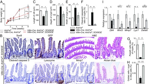
Fig. 5.
Sox9 overexpression rescues growth failure, abnormal intestinal structure, and skewed differentiation in intestinal Arid1a mutant mice. (A) Body weight at indicated time points for Arid1af/f (black dashed line, n = 6), Villin-Cre;Arid1af/+;SOX9OE (red dashed line n = 3), Villin-Cre;Arid1af/f (black line, n = 6), and Villin-Cre;Arid1af/f;SOX9OE mice (red line, n = 5). (B) IHC analysis for Sox9, GFP, H&E, cleaved caspase 3, lysozyme, Mmp7, and Alcian blue in Villin-Cre;Arid1af/f;SOX9OE intestines at 8 wk of age. [Scale bars, 50 µm (short) and 100 µm (long).] (Inset magnification, 2.7×.) (C–E) Average length of villi (C), depth of crypts (D), and width of crypts (E) in control, Villin-Cre;Arid1af/f and Villin-Cre;Arid1af/f;SOX9OE mice at 8–10 wk of age (n = 3). (F) Ratio of the number of crypts that contained at least one cleaved caspase 3+ cell to all crypt numbers in sections from control, Villin-Cre;Arid1af/f, and Villin-Cre;Arid1af/f;SOX9OE mice at 8–10 wk of age (n = 3). (G) Ratio of the number of Paneth cells to crypt cells in control, Villin-Cre;Arid1af/f, and Villin-Cre;Arid1af/f;SOX9OE mice at 8–10 wk of age (n = 3). (H) Ratio of the number of Goblet cells to crypt to villus cells in control, Villin-Cre;Arid1af/f, and Villin-Cre;Arid1af/f;SOX9OE mice at 8–10 wk of age (n = 3). (I) Relative expression levels of Paneth cell markers in control, Villin-Cre;Arid1af/f, and Villin-Cre;Arid1af/f;SOX9OE intestines, as determined by q-PCR using crypt RNA at 8 wk of age (n = 5). Quantitative data are presented as means ± SD, *P < 0.05, **P < 0.01, ***P < 0.001.

