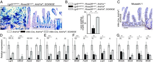
Fig. 7.
Sox9 overexpression restores ISCs in Arid1a mutant mice. (A) Macroscopic images of staining for LacZ and staining for Arid1a and LacZ in the small intestine in Lgr5CreERT2/+;RosalacZ/+;Arid1af/f;SOX9OE mice at 21 d after the last tamoxifen injection. [Scale bar for both panels, 100 µm.] (Inset magnification, 2.7×.) (B) The percentage of crypts containing at least one LacZ+ cell in the control Lgr5CreERT2/+;RosalacZ/+;Arid1a+/+, Lgr5CreERT2/+;RosalacZ/+;Arid1af/f, and Lgr5CreERT2/+;RosalacZ/+;Arid1af/f;SOX9OE mice at 8 wk of age (n = 3). (C) Staining for Musashi-1 in Villin-Cre;Arid1af/f;SOX9OE mice at 8 wk of age. (Scale bar, 100 µm.) (Inset magnification, 2.7×.) (D–F) Relative expression levels of intestinal stem cell markers (D), Wnt target genes (E), and Wnt agonist and receptor genes (F) in control, Villin-Cre;Arid1af/f, and Villin-Cre;Arid1af/f;SOX9OE intestines by q-PCR using crypt RNA at 8 wk of age (n = 5). (G) Relative expression levels of Wnt agonist genes in control, Villin-Cre;Arid1af/f, and Villin-Cre;Arid1af/f;SOX9OE intestines by q-PCR using whole tissue RNA at 8 wk of age (n = 3). Quantitative data are presented as means ± SD, *P < 0.05, **P < 0.01, ***P < 0.001.

