Fig. 2.
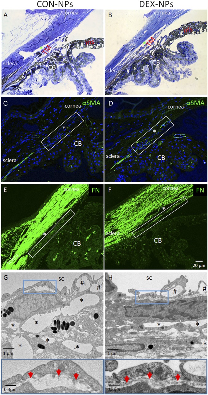
Morphological changes in conventional outflow tissues of mouse eyes treated with DEX-NPs. Age- and gender-matched C57BL/6 mice were injected bilaterally once per week for 2 mo with either vehicle-loaded NPs (CON-NPs) or DEX-loaded NPs (DEX-NPs). (A and B) Iridocorneal angle tissue morphology of CON-NP− versus DEX-NP−injected mouse eyes, visualized by light microscopy after methylene blue staining, showed no gross difference between DEX-NP− and CON-NP−injected eyes. (C–F) Changes in α-SMA and FN levels in iridocorneal angle tissues of mouse eyes injected bilaterally with CON-NPs vs. DEX-NPs by immunofluorescence microscopy. Identical confocal settings were used for experimental and control groups. Conventional outflow tissues are outlined by white box, and nuclei are counterstained with DAPI. (A−F: scale bar, 20-µm.) (G and H) (Top) Ultrastructure of conventional outflow tissues in DEX-NP−treated eyes compared with CON-NP−treated eyes. Giant vacuoles in Schlemm’s canal (SC) are indicated by hash marks. So-called “open spaces” in the juxtacanalicular region of the trabecular meshwork are indicated by asterisks. (Bottom) Boxed areas are enlarged, showing changes in basal lamina beneath SC endothelial cells. Red arrows indicate continuous basement membrane in DEX-NP−treated eyes compared with the discontinuous basal lamina seen in CON-NP−treated eyes (representative images from n = 4 animals); CB, ciliary body; *, SC lumen.

