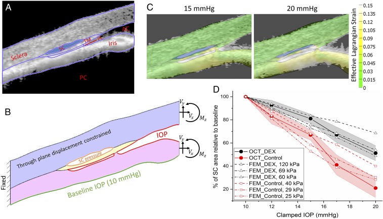
Fig. 4.
Inverse FEM to estimate stiffness of mouse TM. (A) Representative OCT image of a DEX-NP−treated mouse overlain with tissue domains used to construct 2D finite element model. AC, anterior chamber; PC, posterior chamber; PL, pectinate ligament. (B) Resulting finite element model showing locations at which loads were applied, namely the lumen of SC, the anterior chamber, and the posterior iris, as well as the effective loads (Vx, Vy) and moment (Mz) acting on the virtual cut plane at the image boundary, arising from pressures acting on the iris and cornea. (C) Predicted tissue deformations and effective Lagrange strains (color) overlain on corresponding OCT images at 15 and 20 mmHg; note the reasonable agreement between iris position at both pressures. (D) Comparison between experimentally measured SC luminal area at different IOPs, and inverse FEM results, showing an approximately twofold difference in estimated effective TM stiffness (29 kPa in CON-NP−treated eyes vs. 69 kPa in DEX-NP−treated eyes). IOP is set (or clamped) by adjusting the height of a fluid reservoir. The shaded regions show 95% confidence intervals.

