Fig. 1.
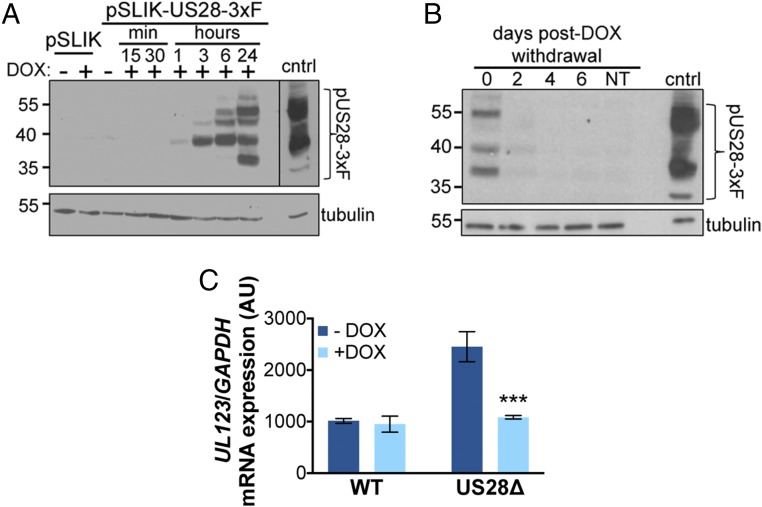
Continual pUS28 expression complements the lytic-like phenotype following US28Δ infection. (A) THP-1 cells were transduced with THP-1-pSLIK-hygro (pSLIK) or THP-1-pSLIK-US28-3xF (pSLIK-US28-3xF) and treated with (+) or without (−) DOX (1 μg/mL) to induce pUS28 expression. Cells were harvested at the indicated time points posttreatment. pUS28 expression was detected by immunoblot using the FLAG epitope tag. (B) THP-1-pSLIK-US28-3xF cells were treated with DOX for 24 h and harvested to confirm pUS28 expression (0 d). Remaining cells were washed in PBS and cultured in the absence of DOX for the duration of the experiment. Cell samples were taken at the indicated days posttreatment and all samples were then immunoblotted for pUS28 using the epitope tag. (A and B) As a control (cntrl), NuFF-1 cells were infected with US28-3xF (moi = 0.5) and cell lysates were harvested at 96 h postinfection. Tubulin is shown as a loading control. Note that due to the intensity of this control lysate in A, a shorter exposure is shown, as denoted by the black line. (C) THP-1-pSLIK-US28-3xF cells were treated without (−; dark blue) or with (+; light blue) DOX for 24 h and then infected with WT or US28Δ (moi = 1.0). DOX was replenished every 48 h and the cells were harvested at 6 dpi for UL123 expression by RT-qPCR. Samples were normalized to GAPDH, and each sample was analyzed in triplicate. Errors bars indicate SD. Statistical significance was calculated using Welch’s t test; ***P < 0.001.

